
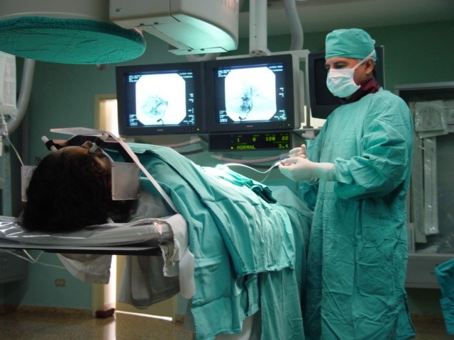

An aneurysm is a weak point in the wall of a cerebral artery that becomes dilated like a balloon and can rupture causing intracranial bleeding. The aneurysm can put pressure on a nerve or surrounding brain tissue. Each bleeding episode carries a high probability of death or disabling neurological sequelae.
The traditional treatment of choice is craniotomy (opening of the skull) and the placement of a clip that meets the objectives of closing the neck at the entrance to the aneurysm sac, and at the same time preserving the lumen or the caliber of the artery that originated the aneurysm. aneurysm.
However, there is a proportion of patients in poor general or neurological condition, as well as aneurysms that could be difficult for the surgeon to locate (for example, aneurysms of the vertebrobasilar circulation). In these cases, an excellent alternative is endovascular embolization techniques. This consists of the introduction of catheters, which are tiny plastic tubes of decreasing calibers, from the femoral artery to the cerebral arteries to be placed inside the same sac of the aneurysm. Once there, we proceed to place platinum coils of calibers adapted to the size of the aneurysmal sac, until it is obliterated. The advantage of this treatment is its minimally invasive condition, without the disadvantages of opening the skull and exposing and manipulating brain tissue. Its disadvantages include that, in the medium to long term, compaction of the skein of coils within the aneurysmal sac is possible, which would lead to recanalization and eventual regrowth of the aneurysm.
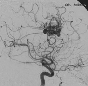
Anatomically, cerebral arteriovenous malformations (AVMs) are a conglomerate of immature and fragile blood vessels that can be located anywhere in the brain, causing a tendency to seizures and neurological deficits on the one hand, and a propensity to produce cerebral hemorrhage on the other.
The best treatment alternative in many cases is craniotomy and microsurgery, however, this is risky for a group of patients with malformations located in eloquent areas of the brain (motor, language, visual, memory, calculation, reasoning, etc.) or very deep brain regions (basal ganglia, midline, brain stem).
For this group of AVMs, there are other treatment alternatives, such as endovascular embolization and stereotactic radiosurgery.

Hemangiomas are benign tumors of the inner layers of blood vessels. The classically recognized hemangioma is a visible red skin lesion that can appear superficially (in the upper layers of the skin, called capillary hemangioma), or at a deeper level (cavernous hemangioma) or a mixture of both. Hemangiomas are usually present at birth, but can appear a few months later and cause a cosmetic problem.
Hemangiomas, both deep and superficial, enter a phase of rapid growth in which their volume and size increase rapidly. This phase is followed by a resting phase, in which the hemangioma changes very little, and by an involution phase in which it begins to disappear.
Hemangiomas can occur anywhere on the body, however, they are more worrisome for parents when they appear on the face or head of their children. Hemangiomas of the eyelid can interfere with the normal development of vision and must be treated within the first few months of life. The size and location of hemangiomas rarely interfere with breathing and eating, so early treatment may be required. Large cavernous hemangiomas may cause secondary infections or ulcerate. Bleeding is usual and can be significant after the hemangioma has been injured.
A good therapeutic alternative is endovascular embolization, which manages to accelerate the involution process of the hemangioma until its disappearance, in a few months.
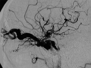
Carotid-cavernous fistulas (CCF) manifest predominantly by red eye, audible murmur, exophthalmos, chemosis, increased intraocular pressure, paralysis of the eye muscles, and loss of visual acuity, among others. When FCC is suspected, it must be promptly confirmed through relevant neuro-ophthalmological and radiological examinations, including an appropriate angiographic study, especially when there is a history of open head trauma.
In the vast majority of FCC, the treatment of choice is endovascular embolization of the fistula site, which should not take long due to the risk of irreversible visual loss and orbital or intracranial hemorrhage caused by long-standing venous hypertension. The embolic agents of choice are detachable balloons and coils. Endovascular interventions should be carried out by appropriately trained and equipped specialists whose experience demonstrates results comparable to contemporary standards, that is, around 95% cure versus 5% complications.
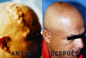
These lesions mainly include arteriovenous malformations and fistulas, as well as venous malformations. Not only are they disfiguring abnormalities, but they can cause devastating, life-threatening bleeding in this group of patients. Appropriate treatment in a significant proportion of cases includes endovascular embolization which could be curative on its own, or used as a first step, to facilitate definitive surgical resection.
A cerebral embolism is the occlusion of a brain artery by a clot formed either in the walls of the heart or in the arteries of the neck and that breaks off traveling to the brain and interrupting the cerebral circulation of a certain area and therefore deprives it. of their nutrition and oxygenation, which leads to the death of nerve cells.
What are the symptoms of a cerebral embolism?
Includes sudden onset of numbness or weakness of one half of the body, confusion, difficulty speaking or understanding language, visual difficulty in one or both eyes, double vision, dizziness, balance disorder.
How should one act in the presence of a possible cerebral embolism?
The patient should be taken immediately to an emergency service, since its immediate treatment could save his life and thus allow the patient to have a rehabilitation that can achieve full recovery or minimize the intensity of the sequelae.
Why is it important to act quickly when dealing with a patient with a cerebral embolism?
Because the embolism patient can receive a drug called r-tPA that dissolves the clots that obstruct the circulation. Ideally, the patient should arrive at a center with vascular neurologists within 1 hour and be evaluated with cerebral tomography and magnetic resonance imaging and treated with appropriate drugs within 3 hours of the onset of the embolic event. In this way, the probability of recovering from the embolism is 30% higher than that of patients not treated with this medication.
What is the prognosis for patients with cerebral embolism?
50% to 70% of the survivors of a cerebral embolism achieve their functional independence but 15% to 30% of them are left with permanent disabling sequelae.
Can a cerebral embolism be repeated?
25% of patients who recover from their first stroke will suffer a new episode within 5 years.
What is a transient ischemic attack (TIA)?
TIA is a neurological malfunction that lasts a few minutes and occurs from a brief interruption of blood flow to a part of the brain. The symptoms of TIA are the same as those of cerebral embolism and most have a maximum duration of one hour although they may persist for a maximum of 24 hours.
Is there treatment for transient ischemic attack (TIA)?
Affirmative. Since there is no way of knowing if the episode of cerebral circulatory failure will be a simple TIA or will progress to a full-blown stroke, specialist evaluation within an hour should determine whether or not clot-dissolving treatment is initiated. In the case of a TIA, the patient must receive antiplatelet drugs permanently and combat risk factors such as obesity, sedentary lifestyle, high blood pressure, increased blood fats, diabetes, smoking, etc Patients with cardiac arrhythmia should receive anticoagulant drugs.
Can clogged arteries in the neck be recanalized?
Definitely. There are two techniques: Endarterectomy or open surgery and endovascular angioplasty placing devices or expandable cylindrical metal meshes that are placed through catheters that are inserted through a puncture in the femoral artery in the groin. Both techniques achieve comparable results and significantly improve the prognosis of patients.
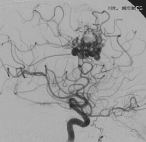
Anatomically, cerebral arteriovenous malformations (AVMs) are a conglomerate of immature and fragile blood vessels that can be located anywhere in the brain, causing a tendency to seizures and neurological deficits on the one hand, and a propensity to produce cerebral hemorrhage on the other.
The best treatment alternative in many cases is craniotomy and microsurgery, however, this is risky for a group of patients with malformations located in eloquent areas of the brain (motor, language, visual, memory, calculation, reasoning, etc.) or very deep brain regions (basal ganglia, midline, brain stem).
For this group of AVMs, there are other treatment alternatives, such as endovascular embolization and stereotactic radiosurgery.
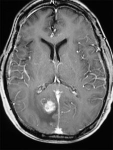
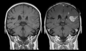
Brain metastases are a fairly advanced manifestation of cancers of other organs of the body. Malignant tumors such as lung, breast, kidney, colon, or skin (melanoma) frequently spread to the brain through the blood and colonize the brain, often with several foci and settle in critical and/or deep areas, therefore an excellent treatment option is radiosurgery.
Radiosurgery has the advantages of radical surgical resection without the disadvantage of the frequent complications associated with craniotomies.
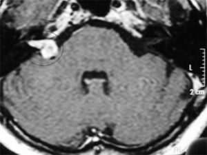
Meningiomas, acoustic neuroma:
After astrocytomas, meningiomas and neurinomas are the most frequent intracranial tumors. Although fortunately they are benign in nature, their intracranial location and their diagnosis, often at an advanced stage, sometimes make them risky lesions for open surgery and resection.
In the case of choosing treatment with open surgery, the embolization intervention prior to surgery manages to reduce intraoperative bleeding and therefore surgery is usually faster and more complete, thus avoiding complications and relapses (See figure))
When open surgery is considered risky, stereotactic radiosurgery is an excellent alternative in these cases, since it is a non-invasive operation, without opening the skull, with the patient awake, and with high-energy radiation, guided by laser. Radiosurgery achieves tumor control at 5 years in about 80% of patients, with a rate of permanent complications equal to or less than approximately 3%.
